The Mammalian Excretory System: Anatomy and Function
Objectives
This blog post provides readers with the
following objectives. The reader will be able to:
EXCRETION IN MAMMALS
Excretion is the removal of the metabolic wastes from the body of an organism. Wastes that are removed include carbon dioxide, water, salt, urea and uric acid.
Excretory Organs in Mammals
1. Liver as an Excretory Organ
1. It excretes bile pigment or bilirubin
2. It disposes worn-out red blood cells or hormones
3. It detoxifies poisonous substances and drug
4. Foreign particles in the blood stream are removed by phagocytic cells in the blood vessels of the liver.
5. It converts excess amino acid or ammonia into urea in a process known as deamination. The urea is eliminated from blood stream through the skin or the kidney.
2. Skin as an Excretory Organ
It removes excess water, salt, urea and uric acid
The sweat glands absorb water, salts and small quantities of urea from the blood. These substances form sweat in the sweat gland which eliminates through the sweat pore.
3. Lung as Excretory Organ
It eliminates carbon dioxide and water vapor, a waste product given off by cellular respiration. These products are removed from the body when expired air diffuses through the nostrils into the atmosphere.
4. Kidneys as Excretory Organ
It filters the blood to form urine, (excess water, salt, urea and uric acid)
Urinary System
It consists of two kidneys, two ureters, the urinary bladder and the urethra.
Structure of the Kidney: Anatomy and Function
The kidneys are essential organs in the human body responsible for filtering blood, removing waste products, and maintaining fluid and electrolyte balance. Understanding their structure is crucial for comprehending their functions and role in overall health.
Overview of Kidney Structure
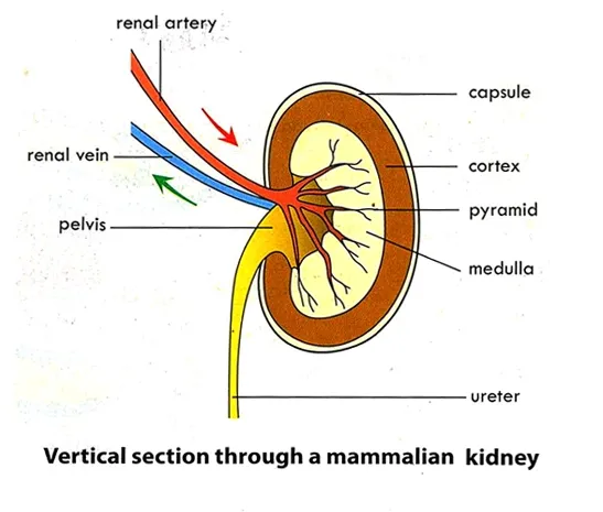
The kidneys are paired, bean-shaped organs located in the lower back region, on either side of the spine. Each kidney is about 10-12 cm in length and is protected by the rib cage and a layer of fat. Kidneys are reddish-brown, bean-shaped organs located behind the peritoneum. Each kidney is cover by outer fatty fibrous layers called capsule. A cone-shaped, adrenal gland lies on top each kidney. Blood vessels, renal artery enters the kidney and renal vein leaves the kidney through a depression termed the hilum.
The hilum also give rise to a long narrow tube called ureter that connects the kidney to an oval, transparent sac called urinary bladder. The longitudinal section of the kidney shows two distinct regions.
Major Structures of the Kidney
1. Renal Cortex
- Description: The outer layer of the kidney, just beneath the renal capsule. It is rich in blood vessels and contains the renal corpuscles, which are the starting points of the nephron.
- Function: Houses the glomeruli, where blood filtration begins.
2. Renal Medulla
- Description: The inner region of the kidney, organized into pyramid-shaped structures called renal pyramids.
- Function: Contains the loops of Henle and collecting ducts that help concentrate urine and reabsorb water and electrolytes.
3. Renal Pyramids
- Description: Cone-shaped structures within the renal medulla, arranged in a radial pattern.
- Function: The sites where urine is collected before being transported to the renal pelvis.
4. Renal Pelvis
- Description: The funnel-shaped structure located in the center of the kidney, where urine collects before entering the ureter.
- Function: Acts as a conduit for urine from the renal pyramids to the ureter.
5. Renal Capsule
- Description: A tough, fibrous outer layer that encloses the kidney.
- Function: Provides protection and helps maintain the shape of the kidney.
6. Nephron
- Description: The functional unit of the kidney, each kidney contains about 1 million nephrons.
- Function: Performs the critical functions of filtration, reabsorption, and secretion to produce urine.
- Components:
- Glomerulus: A network of capillaries where blood filtration starts.
- Bowman’s Capsule: Surrounds the glomerulus and collects filtrate.
- Proximal Convoluted Tubule (PCT): Reabsorbs nutrients, ions, and water.
- Loop of Henle: Creates a concentration gradient in the medulla.
- Distal Convoluted Tubule (DCT): Further adjusts the composition of urine.
- Collecting Ducts: Transport urine to the renal pelvis.
Blood Supply of the Kidney
1. Renal Artery
- Description: Carries oxygenated blood from the aorta to the kidney.
- Function: Supplies the kidneys with blood for filtration.
2. Renal Vein
- Description: Carries deoxygenated blood from the kidney to the inferior vena cava.
- Function: Transports filtered blood away from the kidneys.
Functions of the Kidney
1. Filtration
- The kidneys filter waste products and excess substances from the blood to form urine.
2. Regulation
- They regulate blood pressure, electrolyte balance, and fluid levels in the body.
3. Hormone Production
- The kidneys produce hormones such as erythropoietin, which stimulates red blood cell production.
4. Acid-Base Balance
- They maintain the pH balance of the blood by excreting hydrogen ions and reabsorbing bicarbonate.
For more detailed information on kidney anatomy and function, visit these resources:
- National Kidney Foundation: Kidney Anatomy
- Mayo Clinic: Kidney Function and Anatomy
- Khan Academy: Structure of the Kidney
Structure of the Nephron: Anatomy and Function
The nephron is the fundamental functional unit of the kidney, crucial for urine formation and maintaining fluid and electrolyte balance. Each kidney contains over a million nephrons, which work together to filter blood, reabsorb essential nutrients, and excrete waste products. Here's a detailed look at the structure of the nephron and its components.
Overview of the Nephron
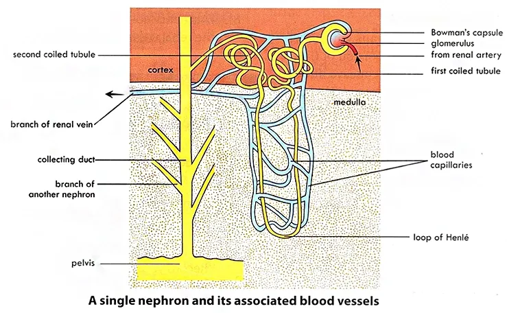
The nephron comprises a network of tubules and associated blood vessels that collectively perform the essential functions of filtration, reabsorption, and secretion. The primary segments of a nephron include the Bowman’s capsule, proximal convoluted tubule, loop of Henle, and distal convoluted tubule.
Components of the Nephron
1. Bowman’s Capsule
- Description: A cup-shaped structure that encases a network of blood capillaries called the glomerulus.
- Location: Located in the renal cortex of the kidney.
- Function: The Bowman’s capsule filters blood plasma into the renal tubule. The glomerulus within the capsule performs the initial filtration of blood.
2. Proximal Convoluted Tubule (PCT)
- Description: A coiled segment of the nephron tubule that follows the Bowman’s capsule.
- Structure: The walls of the PCT are lined with microvilli, which significantly increase the surface area for nutrient reabsorption.
- Function: The PCT is responsible for the reabsorption of water, ions, and nutrients from the filtrate back into the bloodstream.
3. Loop of Henle
- Description: A U-shaped segment of the nephron tubule that extends into the renal medulla.
- Function: The loop of Henle plays a crucial role in creating a concentration gradient in the medulla, which helps in the reabsorption of water and salts.
4. Distal Convoluted Tubule (DCT)
- Description: A coiled segment following the loop of Henle.
- Structure: The DCT is shorter and has fewer microvilli compared to the PCT.
- Function: The DCT is involved in the regulation of potassium, sodium, and calcium levels. It also plays a role in the secretion of hydrogen ions and other substances into the filtrate.
5. Collecting Duct
- Description: The final segment of the nephron where urine is collected from multiple nephrons.
- Function: The collecting duct plays a crucial role in the final adjustment of urine composition and volume by reabsorbing water and sodium.
Function of the Nephron
1. Filtration
- The nephron filters blood to remove waste products and excess substances, forming a filtrate in the Bowman’s capsule.
2. Reabsorption
- Essential nutrients, ions, and water are reabsorbed back into the bloodstream from the filtrate as it passes through the PCT, loop of Henle, and DCT.
3. Secretion
- Additional waste products and ions are secreted into the filtrate from the blood in the DCT.
4. Urine Formation
- The final filtrate, now urine, is collected in the collecting ducts and transported to the renal pelvis for excretion.
For more detailed information about the nephron and its role in kidney function, visit these resources:
- National Kidney Foundation: Structure of the Nephron
- Khan Academy: Nephron Structure and Function
- Mayo Clinic: Nephron Overview
Formation of Urine
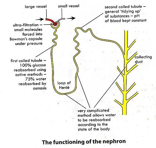
There are three phases in the formation of urine: Ultrafiltration, Selective reabsorption and Tubular Secretion.
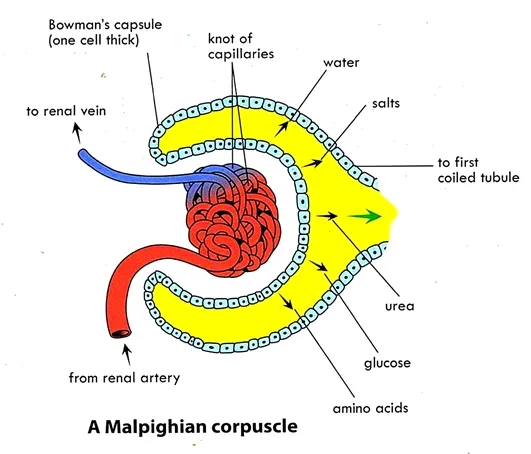
Blood enters the kidney by renal artery. Afferent vessels enter
Bowman’s capsule and form a network of capillaries called glomerulus. The afferent
arterioles of the glomerulus have a greater diameter than the efferent
arterioles. This creates high hydrostatic pressure in the glomerulus which
forces water and certain substance to pass (or filter) into the Bowman’s
capsule. This process is called ultrafiltration
or glomerular
filtration. The filtrate contains small substances such as water,
mineral salts, glucose, amino acids, hormones, urea, uric acid, toxins, drugs
etc.
The non-filterable components such as blood cells, and proteins leave the glomerulus by way of the efferent arteriole. Filtrate that enters the glomerular capsule passes into the lumen of the proximal convoluted tubule.
Selective reabsorption occurs in the
proximal convoluted tubule. Glucose, vitamins, important ions and most amino
acids are reabsorbed from the tubule back into the capillaries surrounding the
tubule. At the loop of Henle, reabsorption of water, sodium ions and chloride
ions take place. At the distal convoluted tubule, there is reabsorption of
salts and water into the surrounding network capillaries. Some wastes are
actively secreted into the fluid by a process called tubular secretion. Some
of these are H+, K+, NH4+ toxic substances and
foreign substances (drugs, penicillin, and uric acid). Secretion of H+
adjusts the pH of the blood.
As fluid passes through the collecting ducts, much of the water reabsorbed by osmosis. The permeability of the collecting duct to water is regulated by Antidiuretic hormone (ADH).
Composition of Urine
Urine contains: water – 96-97%, Urea – 2% and remaining 2% constitute uric acid, creatine, ammonia sodium, potassium, chloride, phosphate, sulphate and oxalates.
Integumentary System
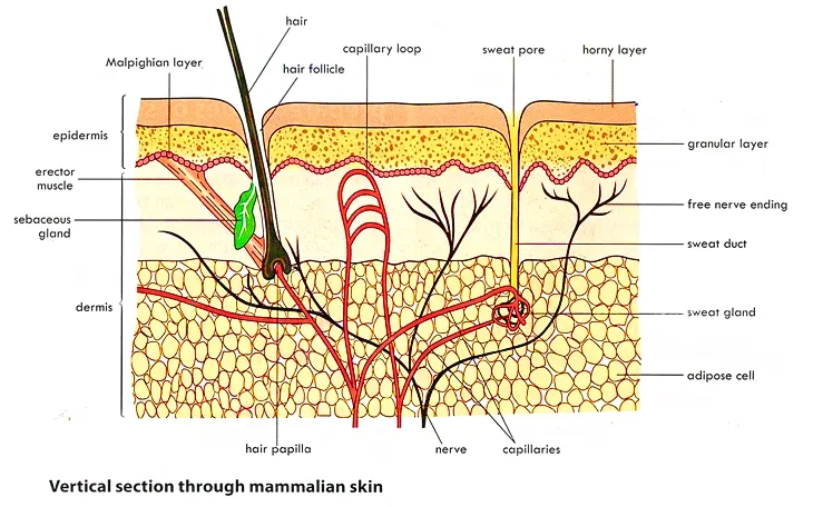
The integumentary system consists of the skin, hair, nails and cutaneous glands.
Structure of Mammalian Skin: Anatomy and Functions
Mammalian skin is a complex and multifunctional organ that serves as a protective barrier, regulates temperature, and allows for sensory perception. It is composed of multiple layers, each with distinct structures and functions. Here’s a detailed overview of the structure of mammalian skin:
Overview of Mammalian Skin
Mammalian skin is composed of three primary layers:
- Epidermis
- Dermis
- Hypodermis (Subcutaneous Tissue)
1. Epidermis
The epidermis is the superficial protective layer of the skin. It is composed of stratified squamous epithelium. It consists of three regions: Malpighian, granular and cornified layer.
1. Malpighian or basal layer: is composed of a single layer of cells in contact with the dermis. This layer composed specialized cells which constantly divide mitotically and replaced those lost from the skin surface. These specialized cells include;
a. Keratinocytes: are cells that produce keratin. Keratin is responsible for the toughness and waterproof covering of the skin.
b. Melanocytes: are epithelial cells that synthesize the pigment called melanin. Melanin provides a protective barrier to the ultraviolent radiation in sunlight. It’s also responsible for skin color.
c. Langerhans cells: are microphages that protect the skin against infection.
2. Granular layer: The granular layer consists of only three or four flattened rows of cells. These cells are produced by the Malpighian layers.
3. Horn-like layer or Cornified layer: This layer is composed of several layers of flattened, cornified, dead cells. These cells constantly shed from the skin surface and replaced by new ones from granular layer. It is the layer that actually protects the skin.
Link: NCBI: Epidermis Overview
2. Dermis
Description: The middle layer of the skin, located beneath the epidermis. It provides structural support and houses various skin appendages. The dermis is deeper and thicker than the epidermis. The dermis contains many sweat glands, oil secreting glands, nerve ending, blood vessels, erector muscles, pigment cells and hair follicles.
1. Hairs: These are formed in tiny pits in the skin called hair follicles. At the base of the follicle there is a cluster of cells called the bulb. The hair is formed by the multiplication of cells of the bulb. The part of the hair above the skin is the shaft and the remainder the root. The hair is responsible for temperature regulation of the skin.
2. Erector muscles: are little bundles of involuntary muscle fibres attached to the hair follicles. Contraction makes the hair stand erect and raise the skin around the hair.
3. Sebaceous glands: are oil-producing glands that empty their secretions into hair follicles. This oily secretion is called sebum. Sebum lubricates and keeps the hair and skin soft. It also inhibits the growth of some bacteria.
4. Sweat glands: secrete sweat onto the surface of the skin. Sweat is composed of water, salts, urea and uric acid. It serves not only for evaporative, but also excretion of wastes.
5. Mammary glands: are found in the breast of female mammals. They are specialized sweat glands that secrete milk during lactation.
Link: Encyclopedia of Dermatology: Dermis Structure
3. Hypodermis (Subcutaneous Tissue)
Description: The deepest layer of the skin, located beneath the dermis. It connects the skin to underlying tissues and organs. It is connective tissues composed of adipose cells. This layer stores fats, insulates and cushions the body and regulates temperature.
Components:
- Adipose Tissue: Provides insulation and stores energy.
- Loose Connective Tissue: Helps anchor the skin to the underlying structures.
- Blood Vessels: Supply nutrients and assist in temperature regulation.
Functions:
- Insulation: Helps maintain body temperature by providing thermal insulation.
- Cushioning: Protects underlying organs and tissues from mechanical damage.
Functions of Mammalian Skin
1. Protection
a. It prevents invasion of micro-organismsb. It protects the body from excessive water lossc. Bacterial are killed by bactericidal substances in sebumd. Melanin absorbs ultraviolent light and protects underlying structures
2. Excretion: the sweat glands excrete salt, water, urea and uric acid in sweat
3. Synthesis: the skin synthesizes vitamin D, which helps regulate calcium and phosphorous metabolism.
4. Sensory Detection: it contains sensory receptors that detect heat, cold, touch and pain.
5. Regulation of Body Temperature
a. The thin layer of fat cells in the dermis insulates the body.
b. Sweat produced by sweat glands evaporate using excess body heat, which cools the body..
For further information on the structure and functions of mammalian skin, consider these resources:
- National Institute of Arthritis and Musculoskeletal and Skin Diseases (NIAMS): Skin Anatomy
- Khan Academy: Skin Structure and Function
- Mayo Clinic: Skin Function and Structure
{nextPage}
Homeostasis
Homeostasis is the maintenance of a constant internal environment in a living organism. It involves a series of automatic control mechanisms.
Examples of homeostasis
1. Regulation of water
2. Regulation of temperature
Significance of homeostasis
1. Maintains a constant internal environment
2. It enables the biochemical process in the body to take place.
Thermoregulation (Temperature Regulation)
Increase in Body Temperature:
An increase in body temperature, for example due to
vigorous exercise or being in a warm place, causes the blood and the skin temperature
to increase. Thermo-receptors in the
skin detect this increase and transmitted a signal in the form of nerve impulse
to the hypothalamus in the brain. When the hypothalamus is stimulated, it then
transmits nerve impulses to the relevant body parts.
1. The arterioles in the skin dilate (a process called vasodilation). More blood is transported to the blood capillaries and this removes more heat.
2. Sweat glands become active and more sweating occurs and hence more heat removed.
3. The hair erector muscles in the skin relax causing the hair to lie flat. Therefore, less heat is trapped by the hair.
4. Metabolic rate decreases and hence heat production decreases.
5. Breathing rate increases so as to remove more heat in exhaled air.
All these mechanisms lower down the blood temperature (Negative feedback) and eventually the temperature will return to normal.
Decrease in Body Temperature:
A decrease in body temperature, for example due to
starvation, fasting or being in cold places, causes the blood and the skin temperature
to decrease. The temperature
receptors in the skin detect this decrease. A signal in the form of nerve
impulse is then transmitted to the hypothalamus in the brain. When the
hypothalamus is stimulated, it then transmits nerve impulses to the relevant
body parts.
1. The arterioles in the skin constrict (a process called vasoconstriction). Less blood is transported to the blood capillaries and hence less heat is removed.
2. Sweat glands become inactive and no sweating occurs. Therefore, no heat is removed.
3. The hair erector muscles in the skin contract causing the hair to stand erect. Therefore, more heat is trapped by the hair.
4. Metabolic rate increases and hence heat production increases.
5. Breathing rate decreases so as to conserve more heat.
6. Shivering due to muscle contractions occurs if very cold to produce more heat.
All these mechanisms increase the blood temperature (Negative feedback) and eventually the temperature will return to normal.
Heat production
Heat is gained by the body cell in following ways
1. The muscle contraction produces large amount of heat. Shivering involves muscles contraction and produces heat.
2. The liver is very chemically active, and heat is produced as a byproduct.
3. The skin absorbs heat directly from the sun.
4. The metabolic reactions of the body produce heat.
Heat loss
1. Most of the heat is loss from the blood through the skin in the form of sweat
2. Small amounts are lost in expire air, urine and faces
3. Heat lost through the skin can only be regulated to maintain a constant body temperature.
Water Regulation (Osmoregulation)
Regulation of the amount of water in the body is controlled by the kidneys and hormone called Antidiuretic Hormone (ADH)
ADH increases the permeability of the distal convoluted tubule and collecting duct. At high osmotic blood pressure (i.e. more salty, not enough water), the posterior pituitary releases ADH. ADH increases the permeability of the collecting ducts, allowing water to leave by osmosis into the blood.
If osmotic pressure of blood decreases, pituitary does not release ADH and more water is lost in urine due to decreased permeability of the collecting duct.
Alcohol inhibits the secretion of ADH, thus increases water loss. The absence of secretion of ADH result in a condition called Diabetes insipidus.
Regulation of Body Sugar
The liver regulates glucose level of the blood. The liver converts excess glucose to glycogen, using insulin produced by pancreatic cells (islet of langerhans), for storage in the muscle or liver (i.e. decreasing blood sugar level).
The hormone glucagon produced by islet of langerhans, promotes the breakdown of glycogen in the liver into glucose in the blood. It also promotes gluconeogenesis, the synthesis of glucose from amino acids or fatty acids (i.e. increasing blood sugar level).
Role of Skin in Homeostasis
The thermoreceptors in the hypothalamus of the brain detect changes in the body temperature and initiate mechanisms to bring the temperature to normal by sending impulses to the erector muscles, sweat gland, blood vessels (arterioles) in the surface of the skin and skeletal muscles.
When the body temperature is low (cold
weather),
1. Erector muscles contract, thereby raising the hair and trapping a thick insulating layer of air which reduces heat loss.
2. Sweat glands produces no or little sweat to reduce or conserve the heat through sweat.
3. Arterioles in the surface of the skin constrict, to reduce the amount of warm blood flowing near to the skin surface, thereby reducing heat loss through radiation.
4. Shivering caused by spontaneous skeletal muscles contraction produces heat which raises the body temperature.
When the body temperature is high (hot weather)
1. Erector muscles of the hair relax making the hair to lie flat, leaving a thin blanket of air so that heat is lost readily from the body.
2. Sweat glands produce sweat and heat is lost through evaporation of the sweat.
4. Arterioles in the surface of the skin dilates, causing more warm blood to be brought near the skin surface for heat to be lost through radiation.
5. Shivering stops and the muscles generate less heat.
Role of Kidney in Homeostasis
When the amount of water in the body is low, the osmotic pressure of the blood rises due to high salt concentration. This change is detected by osmoreceptors in the hypothalamus of the brain. The hypothalamus sends nerve impulse to the pituitary gland to release more ADH (antidiuretic hormones or vasopressin) into circulation. The ADH increase the permeability of the collecting ducts and more water is reabsorbed. On the other hand, when the amount of water in the body is more, the osmotic pressure of the blood reduces, the hypothalamus detects this change and less ADH is released. The collecting ducts become less permeable and less water is reabsorbed.
Role of Lung in Homeostasis
The lung maintained homeostasis in two ways: gas exchange and regulation of blood pH. The lungs eliminate carbon dioxide, a waste product given off by cellular respiration. As carbon dioxide exits the body, oxygen needed for cellular respiration enters the body through the lungs. The removal of carbon dioxide gives the blood a more neutral pH, allowing hemoglobin to bind up more oxygen.
Role of Liver in
Homeostasis
1. The liver controls the blood sugar levels. (N/B: refer to sugar regulation for detail)
2. It detoxifies many poisonous substances, such as some drugs, ammonia, alcohol etc.
3. It removes foreign particles in the blood stream by phagocytic cells in its blood vessels.
4. It converts excess amino acid or ammonia into urea in a process known as deamination.
5. It stores irons from breakdown of red blood cells which is reused to from hemoglobin.
6. The liver also produces heat resulting from its excessive metabolic activities.
7. It stores fats which is released when the need arises.
Get Your Free PDF!
Download our comprehensive guide to Cell Biology to boost your knowledge.
Click the button above to get instant access to the PDF.
Related Post on Biology Topics
Click Here for WAEC Past Questions and Answers on Excretion in Mammals


