CIRCULATORY SYSTEM IN HUMAN
This blog post provides readers with the following objectives. The reader will be able to:
o Explain the concept “Transport” and its need in mammals.o Describe the structure of the mammalian heart.o Explain the mechanism of heart excitation and contractions.o Describe the structure of blood vessels.o Describe the composition of blood.o State the functions of blood.o Describe circulation of blood of a mammal.o Explain the formation of lymph.o Outline the functions of lymph.
CIRCULATORY SYSTEM
The circulatory system consists of two main divisions;
¨ Cardiovascular System: consists of the heart and the blood vessels which pump and carry blood respectively.
¨ Lymphatic System: Consists of the lymphatic vessels and lymphoid tissues within the spleen thymus, tonsils and lymph nodes.
Types of Circulatory System
Open Circulatory System
In an open circulatory system, blood is pumped from the heart through blood body cavities, where it flows over tissues. Blood flows slowly because there is little blood pressure. Arthropods and most mollusks have an open circulatory system.
Closed Circulatory System
In a closed circulatory system, blood is contained within blood vessels, pressure is high, and blood is therefore pumped faster. Valves prevent the backflow of blood within the blood vessels. This type of circulation is found in vertebrates and some invertebrates such as annelids, squids.
BLOOD
Blood is a fluid connective tissue consisting of cells and plasma. It carries oxygen and nutrients to and waste materials away from all body tissues. It is slightly alkaline, with a pH between 7.35 – 7.45. Blood accounts for approximately 8% of the body weight.
Functions of the Blood
The functions of the blood are categories into transport, protection and regulation.
Transport
o carries oxygen from the lungs to other organs
o carries nutrients from the digestive system to all parts of the body
o carries carbon dioxide from cells to the lungs for removal
o carries hormones from the endocrine glands to their target cells
o carries other wastes to the liver and kidneys for detoxification and excretion
o carries metabolic heat to the skin for removal; and help to stabilize body heat
o carries platelets and blood proteins
Protection
o Leukocytes destroy some microorganisms and cancerous cells.
o Antibodies and complement proteins neutralize toxins or help to destroy microorganisms.
o Clotting of blood to minimize excessive blood loss.
Regulation
o Blood transfers water to and from body tissues which help to stabilize water content.
o Regulation of acid-base balance: Blood helps to stabilize body pH (7.35-7.45).
o Regulate body temperature; warm blood is transported from the interior to the surface of the body, where heat is released from the blood.
Components of Blood
Blood is the only fluid tissue in the body. It has both Cellular and liquid components. That is, the blood consists of living blood cells suspended in a nonliving fluid matrix called plasma.
Types of blood cells (Cellular Components)
o Erythrocytes (Red Blood Cells)
o Leukocytes (White Blood Cells)
o Thrombocytes (Platelets)
Plasma
Plasma is the liquid part of blood.
It is a pale yellow fluid that consists of about 91% water and 9% other
substances, such as proteins, ions, nutrients, gases and waste products.
o Plasma proteins: constitute 7% to 9% of the plasma. The three types of proteins are; Albumins, Globulins and Fibrinogen. Other Proteins/Regulatory Substances include metabolic enzymes, antibacterial proteins and hormones.
o Waste Products: such as by-products of cellular metabolism such as urea, uric acid, creatinine and ammonium salts.
o Nutrients (Organic Materials): these are materials absorbed from digestive tract and transported for use throughout the body. They include glucose and other simple carbohydrates, amino acids, fatty acids, glycerol and vitamins.
o Electrolytes/ ions: such as cations and anions. The cations are sodium, potassium, calcium, magnesium. The anions include chloride, sulphate and bicarbonate.
o Gases: include oxygen, carbon dioxide and nitrogen. They are necessary for aerobic respiration; electron-transport chain and also help buffer blood.
Erythrocytes (Red Blood Cells)
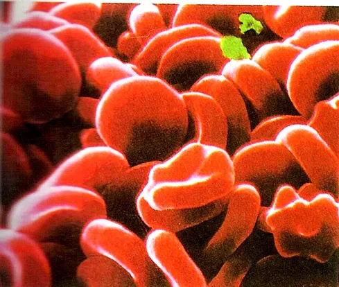
Red blood cell is a small, flattened, biconcave disc shape. It lacks nucleus and mitochondrion. It contains pigmented protein called hemoglobin that binds oxygen. The biconcave shape increases the surface area for more absorption of oxygen.
Absence of nucleus enables the cell to pack more haemoglobin. It also makes the cell more flexible to squeeze through blood capillaries. Red blood cell is manufactured by stem cell in the bone marrow. It has a short life span of about 120 days..
 |
| Scanning electron micrograph of human red blood cells |
Hemolysis of Red blood cell
Red blood cell ruptures (haemolysed) by Kupffer cells of the liver or macrophages in the spleen and bone marrow. Iron in the cell is released and stored in the liver or carried to the bone marrow for the production of new red blood cell.
Liver cells (hepatocytes) pick-up the remaining portion of the hemoglobin, convert into bilirubin and release it in the bile.
Leukocytes (White Blood Cells)
White blood cell is large, irregular shape cell. It contains nuclei, mitochondria and can move in an amoeboid fashion. Amoeboid ability enables the cell to squeeze through pores in capillary walls. White blood cells are classified according to their staining properties. Leukocytes that have granules in the cytoplasm are called granulocytes or granular. Leukocytes without visible granules are called Agranulocytes.
Granulocytes
Granulocytes are large, roughly
spherical and have a short life span.
They have rounded nuclei masses connected by thin nuclear material. Granulocyte
are phagocytes.
Types of granulocytes; neutrophils, basophils and eosinophils.
o Neutrophils: the granules take up both basic (blue) and acidic (red) dyes color. The nuclei consist of three to six lobes. Neutrophils are involved in engulfing and destroying foreign invaders e.g. bacteria. They also secrete lysozymes, enzymes which destroy certain bacteria.
o Eosinophils (Acidiophils): contain granules that stain bright red with eosin, an acidic stain. They produce enzymes that destroy inflammatory chemicals like histamine. They also release toxic chemicals that attack certain worm e.g. tapeworms, flukes, hookworms.
o Basophils: contain large cytoplasmic granules that stain blue or purple with basic dyes. They contain histamine, which release in tissues to increase inflammation. They also release heparin, which inhibits blood clotting.
Agranulocytes
They include lymphocytes and monocytes. They lack visible cytoplasmic granules. The nuclei are spherical or kidney shaped.
o Lymphocytes: are produced by the lymph glands or the lymph nodes. They have large rounded nucleus with small amount of non-granular cytoplasm. Lymphocytes produce antibodies which fight against viruses.
o Monocytes: are largest leukocytes, have kidney shaped nuclei. They are transformed into macrophages and migrate through tissues. Macrophages are phagocytic in nature and they defend the body against viruses and certain intracellular bacterial parasites.
Platelets (Thrombocytes)
Platelets are actually not true cells. They are small fragments of large cells called megakaryocytes. They consist of small amount of cytoplasm surrounded by a plasma membrane. They are ovoid, irregular or disk-like in shape. The cytoplasm contains actin and myosin which cause contraction of platelet. Platelets are produced within the red bone marrow. Platelets play an important role in blood clotting.
Coagulation
When there is cut or wound, coagulation or blood clotting, results in the formation of a clot.
Blood clot: is a network of thread like protein fibers, called fibrin that traps blood cells and fluid. Blood clotting depends on proteins, called coagulation factors, found within plasma. Coagulation factors are in an inactive state and activated by ions to produce a clot.
Mechanism of Blood Clotting
1. At cut or wound, the damaged tissues and blood platelets release an enzyme called thromboplastin also known as tissue factor.
2. The thromboplastin together with calcium ions and vitamin K, convert (inactive) protein prothrombin to active enzyme thrombin.
3. The thrombin then converts soluble protein fibrinogen into insoluble threads of fibrin.
4. The fibrin threads form a mesh or network to trap blood cells to form a clot.
5. White blood cells within the clot fight against invaders at the cut or wound.
{nextPage}
HEART
The heart is a pear-shaped, hollow muscular organ that pumps blood
throughout the blood vessels. It is
located within the middle thoracic cavity. It is enclosed by a double -walled
Sac called the pericardium or pericardial Sac. The pericardium
contains pericardial fluid which
nourishes the heart and prevents shocks. The heart consists of four chambers; two
upper chambers with thin called atria or (auricles) and two lower
chambers with thick walls ventricles. The right side is
separated from the left side by a muscular wall called median septum.
The heart also has valves.
o Tricuspid valve lies between the right atrium and the right ventricle. It consists
of three flaps (hence the name). The flaps are held in position by tendons.
o Bicuspid valve (or mitral valve) with
two flaps lies between the left atrium and the left ventricle. The valves
prevent backflow of blood from the ventricles to the atria.
Mode of Action of the Heart / Cardiac cycle
The cardiac cycle refers to repeated pattern of contraction and relaxation of the heart. The contraction phase is called systole and relaxation phase is called diastole. When the two atria contract the blood is forced into the relaxed ventricles. After a slight pause, the two ventricles contract, forcing the blood into the arteries. The backflow of blood into the atria is prevented by the sudden closing of the tricuspid and the bicuspid valves. The closing of these valves produces a loud “lub” sound which we can hear in a heartbeat.
{nextPage}
Blood Vessels: Types, Functions, and Importance
Overview of Blood Vessels
Blood vessels are a network of tubes that transport blood throughout the body. They are essential components of the circulatory system and play a crucial role in maintaining homeostasis by delivering oxygen and nutrients to tissues and removing waste products. Blood vessels come in different types, each with specific functions and characteristics. There are three main types of blood vessels: arteries, veins and capillaries.
Types of Blood Vessels
Arteries
Arteries are thick, strong, elastic vessels that carry blood away from the heart. Arteries give rise to thinner tubes or branches called arterioles. They have narrow lumen with no valves. Thick wall enables the artery to withstand high pressure of blood from the heart.
o The innermost layer (tunica interna) made of simple epithelium called endothelium. Endothelium prevent blood clotting by provide a smooth surface that allows blood cells and platelets to flow through without being damaged.
o The middle layer (tunica media) has thick layer of smooth muscles and elastic fibers. The fibers enable the arteries to dilate or stretch when the heart pumps blood into them at high pressure.
o The outer layer (tunica externa) is thin and consists of connective tissue with elastic and collagen fibers. This layer attaches the artery to the surrounding tissues.
Examples: The aorta (the largest artery), the carotid arteries (supplying the head and neck), and the coronary arteries (supplying the heart muscle).
Veins
Veins are vessels that carry blood from capillaries back to the heart. Veins have thin wall, less muscular and less elastic. They have wide lumen with valves to prevent backflow of blood. The blood flows with less resistance. Smallest veins are called venules.
They also have three layers:
- Tunica Intima: The innermost layer, lined with endothelial cells and equipped with valves.
- Tunica Media: The middle layer, with less smooth muscle and elastic tissue.
- Tunica Externa: The outer layer, made of connective tissue.
Capillaries
Capillary is small, narrow tube with one-cell-thick walls and narrow lumen. It composed only of endothelium with a basement membrane. Capillaries supply cells with materials and oxygen and take away waste products. Capillaries link the arterioles to the venules within the tissues.
Types:
- Continuous Capillaries: Have uninterrupted endothelial linings, found in most tissues.
- Fenestrated Capillaries: Have pores that allow for greater exchange, found in the kidneys and intestines.
- Sinusoidal Capillaries: Have larger gaps and are found in the liver, spleen, and bone marrow.
Differences between Arteries and Veins
|
Arteries |
Veins |
|
Carry blood away from the heart |
Carry blood towards the heart |
|
Transports blood under high pressure |
Transports blood under lower pressure |
|
Blood flows fast |
Blood flows more slowly and smoothly |
|
Have relatively narrow lumens |
Have relatively wide lumens |
|
Have more muscles/ elastic tissue |
Have relatively less muscles/elastic tissue |
|
Have no semi lunar valves |
Have semi lunar valves to prevent back flow of blood |
|
Carry red oxygenated blood (except: pulmonary arteries) |
Carry bluish-red deoxygenated blood (except: pulmonary veins) |
Functions of Blood Vessels
1. Transport of Blood
- Blood vessels transport blood throughout the body, delivering oxygen and nutrients to cells and removing carbon dioxide and metabolic wastes.
2. Regulation of Blood Pressure
- Arteries and veins regulate blood pressure and flow. Arteries adjust their diameter to manage blood pressure, while veins use valves and muscle contractions to return blood to the heart.
3. Exchange of Substances
- Capillaries facilitate the exchange of gases, nutrients, and waste products between the blood and tissues through their thin walls.
4. Thermoregulation
- Blood vessels help regulate body temperature by adjusting blood flow to the skin. In cold conditions, blood vessels constrict to preserve heat, while in hot conditions, they dilate to release heat.
Disorders of Blood Vessels
Several conditions can affect blood vessels, including:
- Atherosclerosis: A condition where fatty deposits (plaques) build up in the arterial walls, leading to reduced blood flow and increased risk of heart attack or stroke.
- Hypertension (High Blood Pressure): A chronic condition characterized by elevated blood pressure, which can damage blood vessels and increase the risk of cardiovascular diseases.
- Varicose Veins: Swollen, twisted veins usually found in the legs, caused by weakened vein valves and poor blood flow.
- Deep Vein Thrombosis (DVT): The formation of blood clots in deep veins, often in the legs, which can lead to serious complications if the clot travels to the lungs (pulmonary embolism).
Importance of Blood Vessels
Blood vessels are vital for:
- Maintaining Circulatory Health: Efficient blood flow ensures proper oxygen and nutrient delivery to tissues and organs.
- Supporting Metabolic Functions: The exchange of substances at the capillary level is crucial for cellular metabolism and overall health.
- Regulating Blood Pressure and Temperature: Blood vessels play a role in managing blood pressure and body temperature, contributing to homeostasis.
For more detailed information on blood vessels and their functions, visit these resources:
Pathways of Blood Circulation
In mammals, there is a double circulation, i.e. blood passes through the heart twice in one complete circuit. The double circulation consists of two parts:
Pulmonary circulation
From the heart, the pulmonary arteries carry the deoxygenated blood to the lungs. In the lungs the blood gains oxygen and at the same time releases carbon dioxide. The oxygenated blood is then returned to the heart by the pulmonary veins.
Systemic circulation
From the left side of the heart, the oxygenated blood is then distributed by arteries to all parts of the body (except the lungs). The blood releases the oxygen to be used for tissue respiration and at the same time gains carbon dioxide. Deoxygenated blood from body parts is then carried back to the right side of the heart by veins.
{nextPage}
Formation Lymph and Tissue Fluid
The cells in the walls of the blood capillaries do not fit together
exactly. So there are small gaps between them. Plasma and white blood cells
leak out of the blood capillaries. Red blood cells however too large to pass
through these gaps. The fluid formed in this way is called tissue fluid. It surrounds all the cells in the body.
In the tissues, together with the blood capillaries are the lymphatic capillaries. The tissue fluid slowly drains into the lymphatic capillaries. The fluid is now called lymph. The lymphatic capillaries join up to form larger lymphatic vessels. The lymphatic vessels carry the lymph to the sub-clavian veins where it enters the venous blood. The lymph vessels have valves to ensure that movement is only in one direction. Lymph flows much more slowly than blood.
Functions of Lymph
o It supplies cells with oxygen and food nutrients diffuse from the blood
o It also removes waste products from the body cells back into the blood
o Carries pathogens to the lymph nodes to be phagocytized
o Returns tissues fluids to the blood stream
Related Posts
- The Nervous System And Endocrine System In Human
- The Skeletal System And Movement In Mammals
- Excretion In Mammals
- Respiratory System In Human
- Circulatory System In Human
- Nutrition, Dentition And Digestion In Mammals


