INTERNAL STRUCTURES OF PLANT ROOTS, STEMS AND LEAVES
This blog post provides readers with the following objectives. The reader will be able to:
Internal Structure of Root
The longitudinal section through root shows different zones or regions.
Root cap
The outermost protective tip of the root. It protects the delicate apical region. It also eases the movement of the root through soil. It slimy substance, mucigel, which assist the movement of the root tip trough the soil
- Learn more about the Root Cap on Britannica.
Meristematic region or Cell Division)
It consists of meristematic cells. Located just behind the root cap. The cells actively divide to form new cells which differentiate to form specialized root tissues. The new cells replace the worn-out cells of the root cap and also increase the length of the root
- Explore the Meristematic Zone on PubMed Central.
Region of elongation
Cells produced in the meristematic have vacuoles that absorbs water, swells and elongate. This causes the root to elongate. The meristematic and elongation zones are referred to as the region of growth
- For more details, visit Root Elongation Zone on Frontiers in Plant Science.
Region of Maturation or Differentiation
The enlarged cells undergo changes to become the specialized primary tissues. Root hairs, which increase the surface area for water and nutrient absorption, also form in this zone. Primary tissues include epidermis, cortex and vascular system.
- Discover the Zone of Maturation on Britannica.
Primary Tissues of Root
The transverse section of root consists of the following structures
Epidermis
Epidermis is the outer layer of the cells of the young root is responsible for absorbing water and nutrients from the soil.. The cells are closely- packed, thin-walled parenchyma cells with no cuticle, chloroplasts or stomata. The outermost of cells is called piliferous layer. Root hairs arise from the piliferous layer, further enhancing absorption efficiency.
- Learn more about the Epidermis and Root Hairs on PubMed Central.
Cortex
Beneath the epidermis lies the cortex, a layer of parenchyma cells that stores nutrients and helps in the transport of water from the epidermis to the vascular tissue. The cell walls are thickened and contain suberin (fatty substance).The cortex also facilitates gas exchange.
- Explore the Cortex Function on Britannica.
Endodermis
Endodermis forms the innermost layer of the cortex. This single layer of cells surrounds the vascular cylinder and regulates the flow of water and nutrients into the vascular system. The cells are rectangular shape with thickened side walls called casparian strips. The casprin strip is made up of suberin. The casparian strips control the movement of water from cortex into the xylem.
- Discover the Endodermis and Casparian Strip on PubMed Central.
Vascular Cylinder or Stele
Vascular Cylinder or Stele comprises all the tissues enclosed by the endodermis. It consists of the pericycle and vascular tissues (xylem and phloem).
Pericycle
The pericycle is a layer of cells just inside the endodermis. Pericycle is a single cell thick, tightly-packed and meristematic. It gives rise to Lateral roots and vascular cambium.
- Learn about the Pericycle on Britannica.
Vascular tissue or Vascular Cylinder (Stele)
Vascular tissue is conducting tissue in the root. It is central part of the root, and consists of xylem and phloem, which are separated from each other by parenchyma.
□ Xylem: consists of non-living, thick- walled cells. It transports water and dissolved minerals from the roots to the leaves. It also provides structural support for plants.
□ Phloem: alternates between the arms of the xylem and consists of living thin-walled cells. It transports organic substances such as sucrose from the leaves to the roots.
- Explore the Vascular System in Roots on Frontiers in Plant Science.
Functions of Root Structures
- Absorption: Root hairs increase the surface area for water and nutrient absorption. The epidermis plays a key role in taking up these essential resources from the soil.
- Storage: The cortex stores starch and other organic substances that can be utilized by the plant during periods of low photosynthetic activity.
- Transport: The vascular cylinder ensures efficient transport of water, minerals, and nutrients throughout the plant, supporting its growth and development.
- Anchorage: Roots anchor the plant securely in the soil, providing stability and support.
Importance of Healthy Roots
Healthy roots are crucial for overall plant health. They ensure efficient absorption of water and nutrients, support robust growth, and help plants withstand environmental stresses. Maintaining proper soil conditions and avoiding root damage are essential practices for promoting healthy root systems.
Difference between Dicot root and monocot root
|
Monocot Root |
Dicot Root |
|
Xylem is not star-shaped |
Xylem is star-shaped |
|
Wide
pith present |
Pith is absent |
|
Cambium is absent |
Cambium is present |
|
Xylem
has few vessels |
Xylem has many vessels |
|
Secondary growth usually does not occur. |
Secondary growth takes place |
|
Vascular
bundles are many |
Vascular bundles are few |
The Internal Structure of Stems: Key Components and Their Functions
Introduction
The stem is a vital part of a plant's anatomy, serving as the main support structure and transport system. Understanding the internal structure of stems reveals how plants grow, transport nutrients, and adapt to their environments. This article explores the detailed anatomy of stems, highlighting their key components and functions.
A longitudinal section through the stem reveals:
1. Apical meristem or promeristem: the cells at the tip of the stem. The cells constantly divide to produce new cells, which contribute to an increase in length of the shoot.
2. Region of elongation: Cells undergo rapid elongation and enlargement, by taking in water and developing vacuoles, and as result cause growth in length of shoot.
3. Region of maturation: is where the cells of the shoots acquire specific shapes and functions. They are therefore said to be differentiated or specialized.
Key Components of Stem Structure
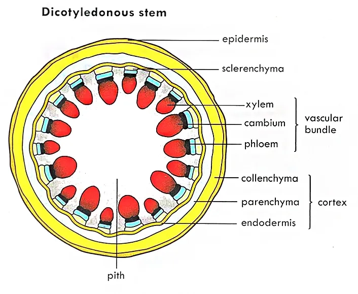
A transverse section through dicot stem reveals the following:
Epidermis
Epidermis is the outermost covering of the stem. It is a single layer of tightly packed, brick-shaped parenchyma cells with no air spaces. There are several unbranched multicellular projections called trichomes. The outer wall is covered with a thin transparent waxy material called cuticle. The cuticle prevents excessive evaporation of water. It also prevents the entry of harmful organisms
- Learn more about the Epidermis on Britannica.
Cortex
It lies next to the epidermis. The
main function is to store food.
It is distinguished into three layers.
1. Collenchyma cells: small cells, the corners are thickened with pectin. This area carries out photosynthesis. It provides mechanical support to the stem.
2. Parenchyma cells: the cells are rounded or angular with intercellular spaces. It provides support to stem due its turgidity.
3. Endodermis: the innermost layer of the cortex. The cells of endodermis are rectangular shaped and closely arranged without intercellular spaces.
- Explore the Cortex Function on Britannica.
Stele (Vascular cylinder)
Stele consist of pericycle, vascular bundles and pith.
Pericycle
Pericycle is the outermost covering of the stele. It’s a layer of compactly arranged sclerenchyma cells. Pericycle strengthens the stem. It also provides protection for the vascular bundles.
Vascular Bundle
Vascular Bundle consists of phloem, xylem, and cambium, arranged in the form of a ring around the central pith..
- Xylem: Transports water and dissolved minerals from the roots to the rest of the plant.
- Phloem: Transports organic compounds, primarily sugars, produced through photosynthesis to various parts of the plant.
- Discover the Vascular System on Frontiers in Plant Science.
a. Cambium
Cambium is present between phloem and xylem. It contains thin walled, brick shaped, meristematic cells i.e., they divide to produce new cells. Cells produced to the outside differentiate as phloem tissue and those to the inside as xylem tissue. The division of the cambium cells results in secondary thickening.
- Learn about the Cambium on Britannica.
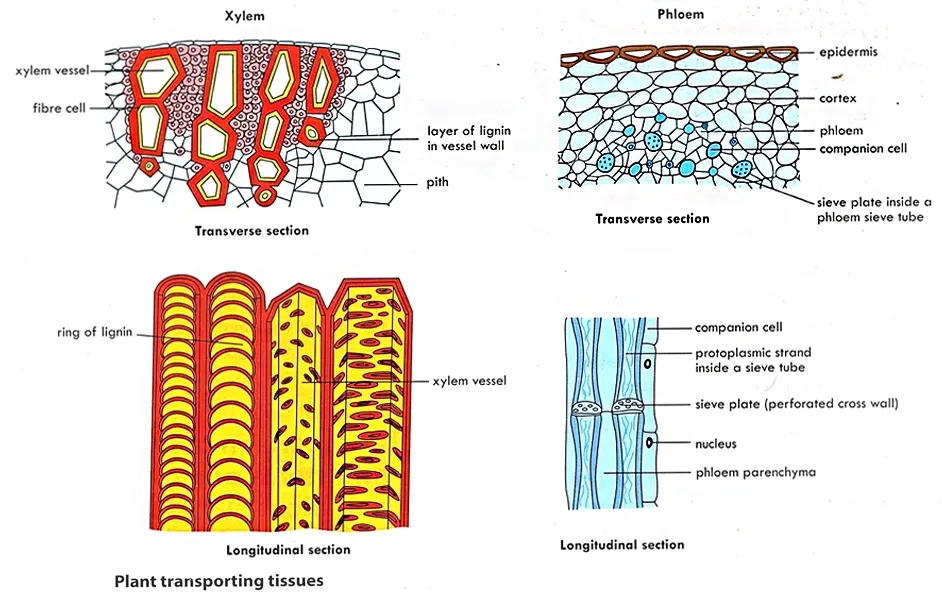
b. Xylem
Xylem transports water and dissolved nutrients from the roots to all parts of the plant. It contains four conducting cells; tracheids, vessel elements, fibers and xylem parenchyma. All the cells have thick lignified walls and are dead at maturity
i. Tracheids: elongated, thin-walled cells with tapered ends. Cells are dead with empty lumen when mature. They have lignified cell walls with their end walls pitted.
ii. Vessel Elements: elongated, hollow tubes arranged end to end. The walls are lignified in variety of ways forming annular, spiral, or reticulate thickenings. The vessel elements transport water more rapidly.
iii. Xylem Fibers: are long cells with much lignified wall. They provide support.
iv. Xylem parenchyma: are living cells, thin-walled, and their cell walls are made up of cellulose. They store metabolites.
c. Phloem
Phloem transports manufacture food throughout the plant. Phloem is composed of sieve tube elements, companion cells and phloem parenchyma.
i. Sieve tube elements: are elongated, tube-like cells, stacked in vertical rows. They are living with no nuclei and have few cytoplasmic organelles. Their end walls are perforated by large pores to form the sieve plates.
ii. Companion cells: are closely associated with sieve tube elements. They are narrow cells with nucleus and dense cytoplasm. They regulate the activities of the sieve tube.
ii. Phloem parenchyma: is made up of elongated, thin-walled cells with dense cytoplasm and nucleus. It stores food material and other substances like resins, latex and mucilage.
Pith
Pith is the innermost part of the stem. It consists of loosely arranged parenchyma cells with intercellular spaces. It stores food and transport nutrients.. n some plants, the pith can also help with the storage of starch. The pith expands among the vascular bundles. These extensions of the pith are referred to as pith rays/medullary rays.
- Explore the Pith Function on Britannica.
Functions of Stem Structures
- Support: The stem provides structural support to elevate leaves, flowers, and fruits, positioning them for optimal light exposure and pollinator access.
- Transport: Vascular tissues in the stem (xylem and phloem) transport water, nutrients, and organic compounds throughout the plant.
- Storage: The cortex and pith store nutrients and water, helping the plant survive periods of drought and nutrient scarcity.
- Growth: The cambium facilitates secondary growth, which increases the girth of the stem and allows the plant to support more leaves and branches.
Types of Stems
Herbaceous Stems: These are soft and green, often found in non-woody plants. Herbaceous stems are flexible and generally do not have secondary growth.
- Learn more about Herbaceous Plants on Britannica.
Woody Stems: Found in trees and shrubs, these stems are hard and have a thick layer of cambium that contributes to secondary growth, forming wood.
- Explore Woody Stems on Britannica.
Climbing Stems: These stems have adaptations that allow them to climb structures for better light access. Examples include vines like ivy and grapes.
- Discover more about Climbing Plants on the Royal Horticultural Society.
Importance of Healthy Stems
Healthy stems are crucial for the overall health and productivity of plants. They ensure efficient transport of water, nutrients, and sugars, support leaves and reproductive structures, and enable plants to compete for light and resources. Ensuring proper care, such as appropriate watering, pruning, and pest control, can maintain stem health.
Difference Between Dicotyledonous Stem and Monocotyledonous Stem
|
Monocotyledonous Stem |
Dicotyledonous Stem |
|
Numerous vascular bundles |
Few vascular bundles |
|
Vascular bundles scattered |
Vascular bundles arranged in a ring |
|
Vascular cambium is absent |
Vascular cambium is present |
|
Pith is absent |
Pith is present |
|
No Secondary thickening |
Secondary thickening occurs |
|
Has medullary rays |
No medullary rays |

The Internal Structure of a Leaf: Key Components and Their Functions
Introduction
Leaves are the primary sites for photosynthesis in plants, converting sunlight into chemical energy. The internal structure of a leaf is intricately designed to optimize this process, along with other vital functions like transpiration and gas exchange. This article delves into the detailed anatomy of leaves, highlighting key components and their specific roles.
Key Components of Leaf Structure
Epidermis
The upper and lower surface of leaf is bound by upper and lower epidermis respectively.
Epidermis is a single layer of rectangular shaped cells containing few or no chloroplasts. The cells are transparent and tightly packed. The epidermis is covered with a waxy, waterproof cuticle. There are a large number of pores called stomata present on the lower surface. Each stomatal pore is surrounded by two bean-shaped cells called guard cells which contain chloroplasts. Stomata controls exchange of gases and transpiration.
- Learn more about the Epidermis on Britannica.
Functions of the Epidermis
1. The cuticle prevents water loss.
2. The epidermis protects the internal tissues from injury.
3. The stomata allows gaseous exchange for photosynthesis and respiration.
4.. The epidermis is transparent which allows light to reach the mesophyll tissue.
Cuticle: The cuticle is a waxy layer that covers the epidermis, preventing water loss and providing a barrier against harmful microorganisms.
- Explore the Cuticle Function on Britannica.
Stomata: These are small openings primarily located on the lower epidermis, surrounded by guard cells. Stomata regulate gas exchange and transpiration.
- Discover the Role of Stomata on PubMed Central.
Mesophyll
Mesophyll is the ground tissue which lies between the upper and lower epidermis. It consists of loosely arranged parenchymal cells. It consist of two distinct regions.
Palisade mesophyll: it occurs below the upper epidermis. It consists of one or more layers of thin-walled, cylindrical cells oriented with their long axis perpendicular to the upper epidermis. The cells are filled with chloroplasts (usually several dozen of them). It is the main site of photosynthesis in the leaf.
Spongy mesophyll: lies beneath the palisade layer. It is made up of loosely packed, irregular shaped cells with large intercellular air spaces. These large intercellular spaces allow free movement of air within the leaf. The air spaces are interconnected and open to the outside through stomatal pores. The cells contain fewer chloroplasts.
Learn about the Mesophyll Structure on Britannica.
Functions of the Mesophyll
The
Veins (Vascular bundles)
The vascular bundles of a leaf extend densely through the mesophyll. Each vein contains two types of vascular tissues: Xylem and Phloem. Veins are surrounded by layers of parenchyma or sclerenchyma cells making up the bundle sheath.
Discover the Vascular System in Leaves on Frontiers in Plant Science.
Functions of the Vascular Bundles
1. The veins strengthen the lamina.
2. The xylem conducts water and dissolved ions to the mesophyll tissue.
3. The phloem conducts organic food from the mesophyll to other parts of the plant.
Cell Types in the Plant
Parenchyma Cells
1. cylindrical in shape and have thin wall
2. living at maturity
3. usually loosely packed, with large intercellular spaces
4. help in synthesizing and storage of synthesized food products
5. controls plant's metabolic reactions
6. play a vital role in healing and regeneration of plants
Collenchyma Cells
1. elongated and irregularly thickened at the corners
2. living at maturity with no intercellular spaces
3. contain few chloroplasts
4. provides a support to herbaceous plants
Sclerenchyma Cells
1. long, narrow and non-living
2. thick, lignified cell walls and lack protoplasts at maturity
3. provide strength and support to plants
4. Sclerenchyma cells are of two types:
i. Sclereids: short cells with irregular shape, thick, lignified secondary walls
ii. Fibers: are long, slender and are arranged in threads.
Differences Between Dicotyledonous and Monocotyledonous Plant
|
Monocotyledonous |
Dicotyledonous |
|
One cotyledon |
Two cotyledons |
|
Narrow
leaf shape, wraps around stem |
Broad, flattened leaf shape |
|
Leaf venation mostly parallel |
Leaf venation mostly net-like or reticulate |
|
Leaves
are sessile (absence of petioles) |
Leaves have petiole |
|
Fibrous root system |
Taproot system |
|
Flower
parts usually in threes or multiples of three |
Flower parts usually in fours or fives or
in multiples of four or five |
|
Usually herbaceous, never woody |
Woody or herbaceous |
|
Pollen
grain has one furrow or aperture |
Pollen grain has 3 furrows or apertures |
|
Vascular bundles in stem are scattered |
Vascular bundles in stem arranged in a ring |
|
Vascular
tissue in root arranged in a ring |
Root xylem usually star-shaped, the phloem
between arms of star |
|
Absence of cambium |
Presence of cambium |
|
Seed
germination is mostly hypogeal |
Seed germination is mostly epigeal |
Download Free Pdf file on Internal Structures of Plant Roots, Stems and leaves
Related Post on Biology Topics
Click Here for WAEC/ SSCE/ WASSCE/ NOVDEC Past Questions and Answers


%20of%20dicotyledonous%20root%20and%20monocotyledonous%20rot.webp)


